

Shear wave elastography of the prostate for benign prostatic hyperplasia and lower urinary tract symptoms
Evaluation of benign prostatic hyperplasia (BPH) and lower urinary tract symptoms (LUTS) rests on symptom questionnaires, uroflowmetry, post-void residual, and anatomic measures of the gland — above all prostate volume. Conceptually, a larger prostate is expected to cause bladder outlet obstruction and the symptoms that follow. In practice, size correlates only weakly with symptoms, and it is uninformative in the men who are symptomatic even though their glands are not enlarged. Size describes only one way for a prostate to obstruct: by bulk.
The histological changes of BPH occur in the transition zone, the tissue immediately surrounding the prostatic urethra. Those changes alter the mechanical properties of that tissue as well as its volume, and stiffer periurethral tissue limits how wide the urethra can open during voiding. Stiffness is therefore a plausible second route to obstruction — but it is not measured in routine care. The existing ways to reach it are invasive: urethral resistance from urodynamic studies (UDS), or mechanical testing of excised tissue.
In this project we propose to measure that stiffness less-invasively, with transrectal shear wave elastography (SWE) of the transition zone. SWE derives tissue stiffness from the speed of shear waves induced acoustically in the tissue, and is already established in breast, liver, and thyroid imaging. Using an endocavitary probe, we sample the tissue immediately around the prostatic urethra, along with the peripheral zone as a reference within the same gland. Our aim is to establish whether size and stiffness are complementary biomarkers describing two routes to obstruction — large, soft glands that obstruct by bulk, and small, stiff ones that do not — and, in the longer run, to identify the men who obstruct without an enlarged prostate, who stand to benefit least from treatments directed at reducing gland volume.
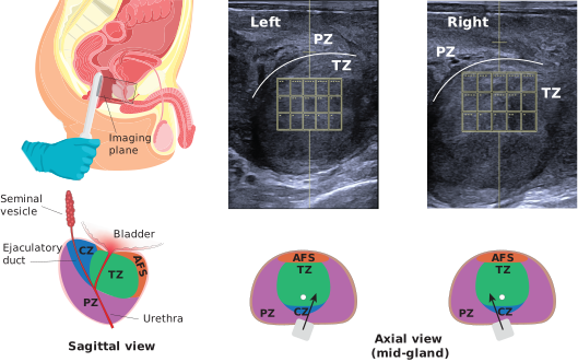
Transrectal SWE of the periurethral transition zone. Left: the endocavitary probe with its imaging plane over the prostate, and the zonal anatomy of the gland — transition zone (TZ), central zone (CZ), peripheral zone (PZ), and anterior fibromuscular stroma (AFS). The TZ surrounds the prostatic urethra and is where BPH arises. Right: ultrasound images of the left and right periurethral TZ, with the grid of sub-regions over which stiffness is sampled, and the corresponding probe orientation in axial view.
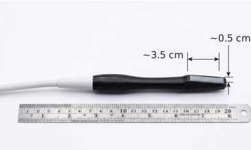
The probe itself: a modified Siemens 18H6 side-firing hockey-stick endocavitary transducer, used with a Siemens Sequoia scanner.
Related Publications
[1] K. Kalayeh, W. Sui, J. Chaffin, M. Zhang, C. L. Brace, M. Grimes, P. Tripathy, T. J. Hall, I. Rosado-Mendez, J. B. Fowlkes and S. Wells, “Shear Wave Elastography Reveals Distinct Prostate Transition Zone Stiffness Across Benign Prostatic Hyperplasia/Lower Urinary Tract Symptoms Phenotypes: A Feasibility Study,” American Institute of Ultrasound in Medicine (AIUM) Annual Meeting, 2026. https://aiumannualconvention2026.eventscribe.net/
[2] K. Kalayeh, W. Sui, M. Zhang, C. L. Brace, M. Grimes, P. Tripathy, T. J. Hall, I. Rosado-Mendez, J. B. Fowlkes and S. Wells, “Prostate Transition Zone Stiffness Measured by Ultrasound Shear Wave Elastography Varies Across Clinical BPH/LUTS Phenotypes: A Feasibility Study,” Annual Meeting of American Urological Association (AUA), 2026. https://www.auanet.org/AUA2026