

Ongoing Projects
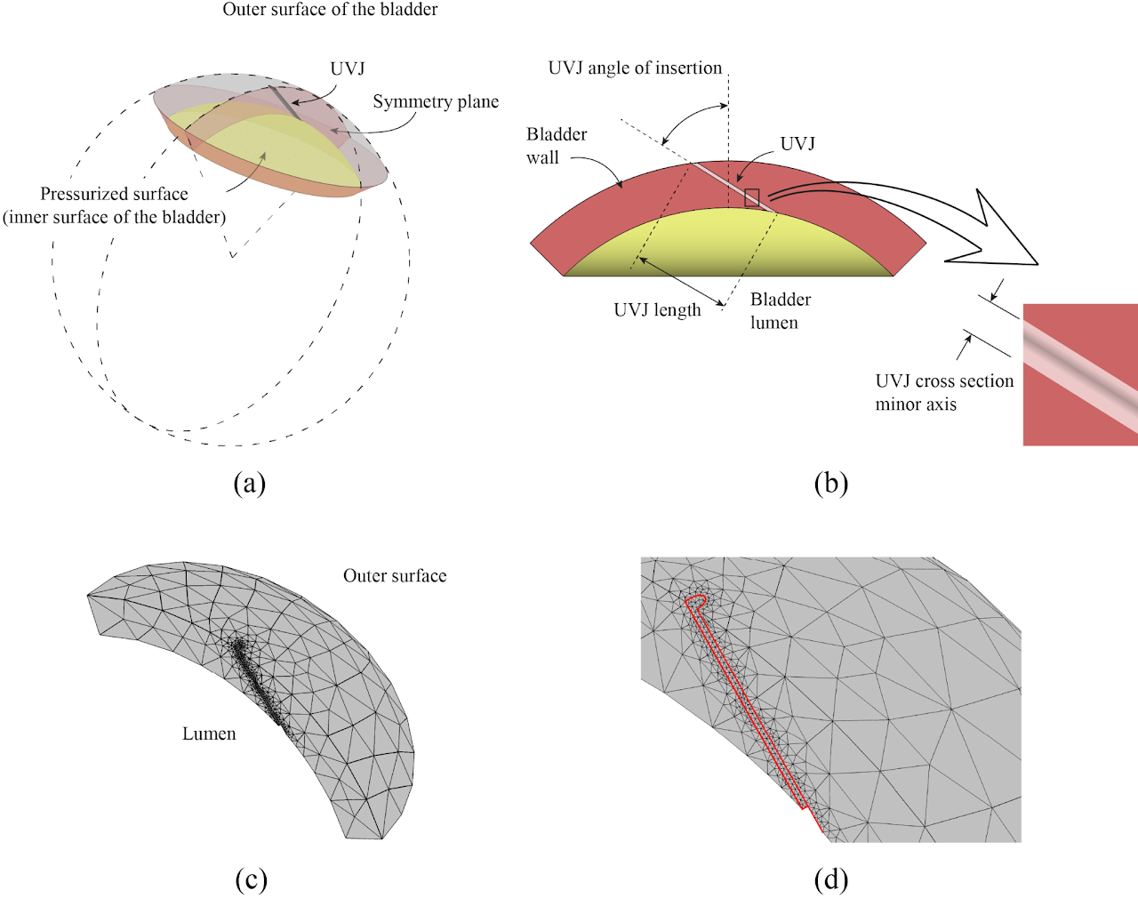
Better understanding of vesicoureteral reflux
In this project, using computational techniques, we set out to better understand the effects of different ureterovesical junction (UVJ) anatomical parameters on vesicoureteral reflux (VUR).
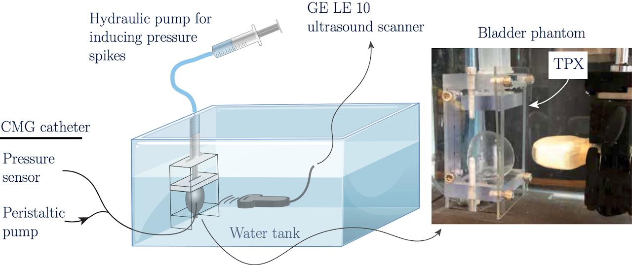
Catheter-free voiding bladder pressure measurement with ultrasound contrast microbubbles
One of the major challenges in urodynamic testing is the presence of an indwelling urethral catheter during voiding pressure evaluation. The space-occupying property of the catheter results in non-physiological conditions, which in turn, can cause misdiagnosis. In this study we hypothesize that ultrasound-based technique known as subharmonic-aided pressure estimation (SHAPE) can measure pressure in a bladder phantom.
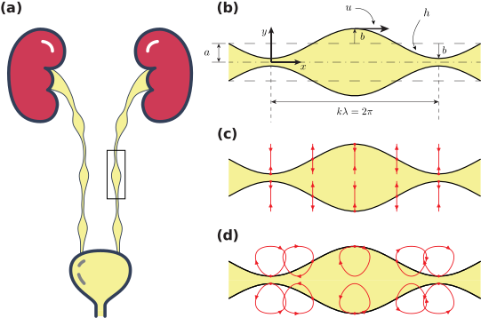
Better understanding of ureteral peristalsis
In this project, we are aiming at better understanding ureteral peristalsis, its normal function and how its abnormal function might cause urinary tract disorders.
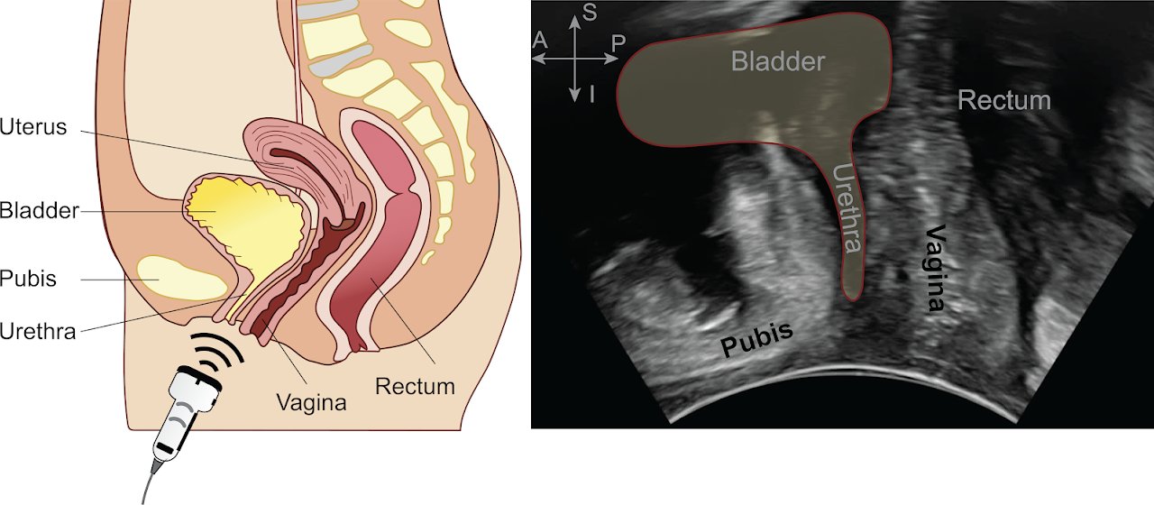
Automated tracking and quantification of pelvic floor motion in transperineal ultrasound for urinary incontinence
Pelvic floor mobility is an important factor in urinary incontinence and transperineal ultrasound can clearly visualize this movement. However, its quantification thus far has been largely a manual process, that focuses on only a small part of the visible movement. In this study, we address a need for an automated methodology to quantify pelvic floor mobility during real-time ultrasound imaging.
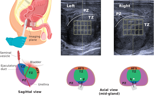
Shear wave elastography of the prostate for benign prostatic hyperplasia and lower urinary tract symptoms
Larger prostates are expected to obstruct the flow of urine, yet gland size correlates only weakly with symptoms and says little about men who are symptomatic without an enlarged prostate. We hypothesize that the prostate obstructs either by bulk or by stiffness, and that the mechanical state of the tissue surrounding the urethra is the part of the picture current evaluation leaves out. In this project we propose to measure that stiffness non-surgically, using transrectal shear wave elastography of the prostate.
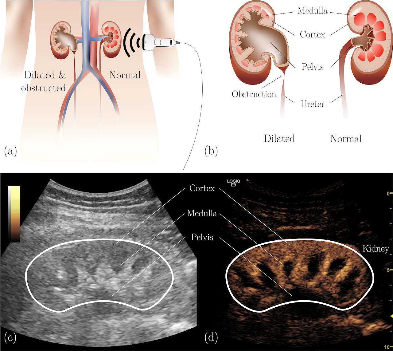
Dynamic contrast-enhanced ultrasound for diagnosing upper urinary tract obstruction
Nuclear medicine diuretic renography (NMDR) is the current standard for diagnosing upper urinary tract obstruction (UUTO), but it is limited by radiation exposure, long imaging times, and reduced sensitivity to early parenchymal changes. This project explores dynamic contrast-enhanced ultrasound (DCEUS) as a safe, fast, and radiation-free alternative that detects microvascular differences between normal and obstructed kidneys.
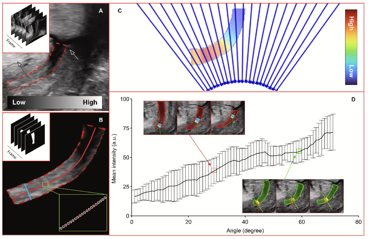
Urethral tissue characterization with angle-dependent multiparametric ultrasound imaging
In this study, we propose to evaluate urethral functionality by assessing its tissue microenvironmental changes with multiparametric ultrasound imaging techniques.
Objective methodology for vesicoureteral reflux evaluation
The current standard method to diagnose vesicoureteral reflux (VUR) is by imaging tests such as voiding cystourethrograms (VCUG) utilizing intravesical infusion of radiographic dye. In such tests VUR is graded based on the visualized degree of dilation and extent to which the contrast flows back up to kidneys. The major limitation of these test is that they ignore ureteral refluxing pressures and volumetric flow rates, which we hypothesize to be crucial for objectivity of these tests and their ability to distinguish between primary and secondary VUR, all important in managing VUR. In this project, we are aiming to develop a new objective methodology to evaluate VUR grade using these biomarkers.
Evaluating pelvic organ prolapse by tracking and quantifying pelvic floor motion in transperineal ultrasound
The amount and timing of different pelvic floor structure movements during various tasks offer important information about pelvic floor motor function. Transperineal ultrasound can clearly visualize this movement in healthy women and those with pelvic organ prolapse. In this study, we address the feasibility of using real-time transperineal ultrasound imaging to perform detailed functional assessments of pelvic floor motor function.
Towards a comprehensive test of urogenital hiatal closure function in women with pelvic organ prolapse
Often injuries during a difficult vaginal delivery result in stretch-related damage to the active and passive tissue of the pelvic floor, resulting in an enlarged urogenital hiatus. However, mechanisms of hiatal closure are poorly understood. In this project, we aim to quantify motor control of hiatal closure in healthy women and women with pelvic organ prolapse using transperineal ultrasound, pressure sensors and surface electromyography.